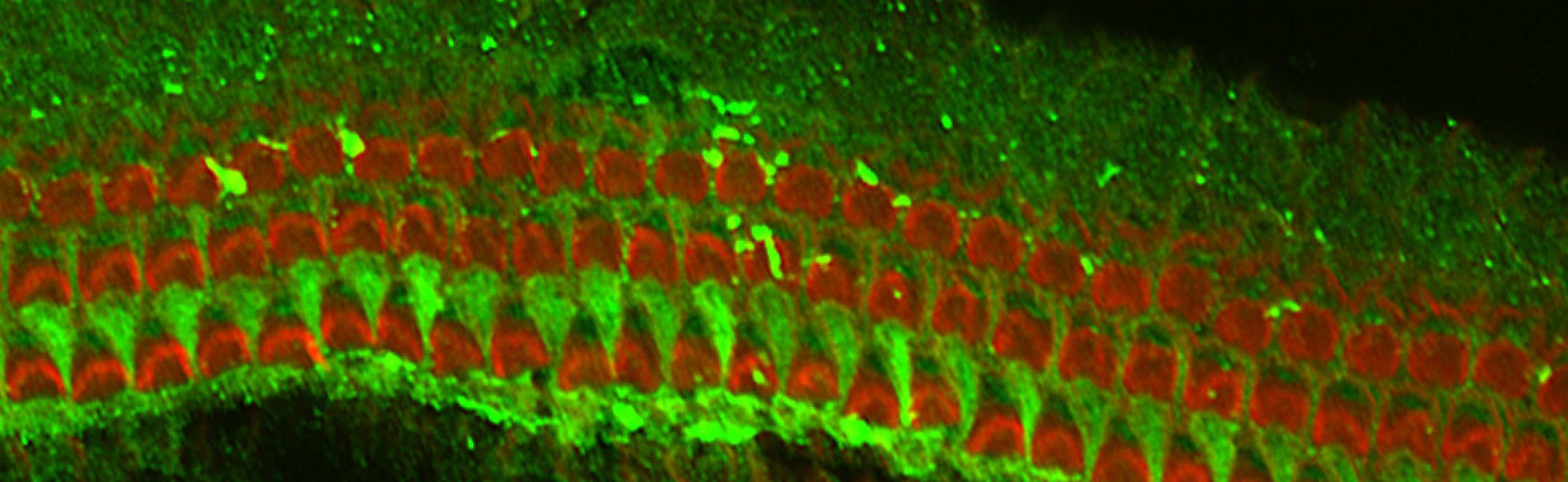
Auditory Brainstem Physiology Laboratory
Research
Descending Neural Connections to the Cochlear Neucleus
Quick Jump:
- The Role of Neural Connections to the Cochlear Nucleus in Sound Coding
- Somatosensory Innervation to the Cochlear Nucleus and its Role in Tinnitus
- The Role of Pathways to the Cochlear Nucleus that are Activated by the Opposite Ear
The Role of Neural Connections to the Cochlear Nucleus in Sound Coding
As in other sensory systems, the cochlear nuclei receive efferent information from higher centers. This is demonstrated especially well in the VCN where most neurons receive extrinsic, non-cochlear synaptic endings from higher auditory and non-auditory centers. In fact, more than half of axosomatic endings on bushy cells in the VCN are non-cochlear, contradicting the commonly held view that these cells are largely relay neurons. A large proportion of their synapses have either flattened or pleomorphic vesicles, usually associated with synaptic inhibition, and contain either glycine or GABA and in some cases both. Large numbers of these putative inhibitory synapses are strategically placed on cell bodies and axon hillocks of bushy and stellate cells to affect the output of these projection.
Sources of non-cochlear innervation arise predominantly in the superior olivary complex, inferior colliculus and contralateral cochlear nucleus (Shore et al, 1991; 1992). Other sources include the cerebral cortex, cuneate nucleus, dorsal column nuclei, interpolar and caudal spinal trigeminal nuclei (Shore et al, 1998; 2000) and vestibular nuclei. Because many of the neurons projecting out of the VCN receive descending information from their target neurons ( Shore et al, 1991; Shore et al, 1992), they may have a feedback function, presumably to enhance or attenuate incoming sensory information from the auditory nerve. We use tract tracing techniques combined with immunocytochemistry to explore the innervation to the cochlear nucleus from non-cochlear sources.
We study the function of these pathways by recording from neurons in the cochlear nucleus. We record from single and multi unit clusters using multichannel electrodes.Another approach to studying the role of these connections is to eliminate them through the use of lesions. We investigate the effects of reversible lesions of the SOC on the spontaneous discharge rate, sound-evoked forward masking functions, responses to clicks and AM sound, and tuning properties of CN neurons. Major sources of CN centrifugal input in guinea pigs are the VNTB, LNTB, MNTB and DMPO and the contralateral cochlear nucleus. Thus, these nuclei are the primary targets for chemical lesions using Lidocaine, Kainic Acid, and Mellitin. (Le Prell et al., 2003).
Somatosensory Innervation to the Cochlear Nucleus and its Role in Tinnitus
Anatomy of Somatosensory Projections to the Cochlear Nucleus
Our recent studies have demonstrated neural pathways from somatosensory regions to the auditory brainstem (Shore et. al., 2000;Zhou and Shore, 2003; 2006a; 2006b, Zhou et al; 2007). The projections terminate in granular and magnocellular regions of the ventral cochlear nucleus (VCN) and surrounding the lateral superior olivary complex (LSO) at the locations of olivocochlear neurons.
These pathways are being studied with tract-tracing and immunocytochemical methods. We have recently identified the transmitter used by the trigeminal nucleus to be glutamate.
Function of the Somatosensory Innervation of the Cochlear Nucleus
We are currently studying function of these projections using electrical stimulation, neuropharmacology and immunocytochemistry. The influence of somatosensory pathways on cochlear nucleus neurons could greatly impact processing in higher auditory centers because a high percentage of information (acoustic and somatosensory) arriving at the CN is conveyed to higher auditory centers.
The project is expedited by the use of multi-channel recording and drug-delivery probes developed by the center for neural communications, enabling us to study the physiological responses of multiple CN neurons simultaneously while delivering drugs or stimulating the trigeminal ganglion. Trigeminal projections to the VCN are mostly excitatory (Shore et. al., 2000, Zhou et al, 2007). Stimulation of trigeminal neurons result in both excitation and inhibition of DCN units. Preliminary studies show this also to be true for neurons in the inferior colliculus.
Role of Somatosensory Innervation of the CN in Tinnitus
Injuries of the head and neck region can lead to the onset of tinnitus in patients with no hearing loss. Furthermore, two thirds of patients with tinnitus (including those with hearing loss) are able to modulate their tinnitus by activating peripheral nerves which innervate the skin or musculature of the face. These observations lead to the hypothesis that somatosensory input to auditory nuclei can play a role in the generation and/modulation of tinnitus. We have shown that the trigeminal ganglion and nucleus innervate the CN and produce changes in neural firing of CN neurons. Therefore, functional connections exists which could explain the observations that head and neck injuries can cause tinnitus and that patients can modulate their tinnitus through somatic activities.
Abnormal activation of nerve fibers, as may occur in injuries of peripheral nerves, can elicit sustained activity in central neurons. In the case of trigeminal ganglion innervation, head and neck injuries could alter the activity of those trigeminal ganglion fibers innervating the CN, resulting in a change in firing rate of CN neurons. These putative "plastic" changes in CNS activity after hearing loss may underly neural hyperactivity which is perceived by patients as tinnitus.
The Role of Pathways Activated by Sound in the Opposite Ear
Additional studies target the function of the cochlear nucleus commissural projection (Shore et al, 1992; 2003), and descending projections from the superior olivary complex and inferior colliculus (Shore et al, 1991, 1992; Shore and Moore, 1998). We stimulate the contralateral ear with sound and assess the effects on spontaneous and sound-driven responses in the ipsilateral cochlear nucleus. The goal of these studies is twofold: a) to determine the function of this pathway and b) to determine whether the ventral cochlear nucleus, by virtue of its binaural properties, plays a role in sound localization. We have shown that contralateral stimuli can suppress/inhibit the activity of VCN neurons, and in a very small percentage of neuorns, activate CN neurons (Shore et al., 2003). Recent studies have shown that this balance of inhibition/excitation can change with deafness. In fact, conductive hearing loss can produce a large amount of excitation by stimulation of the ear opposite to the hearing loss (Sumner et al., 2003). A new focus of our studies is to examine the mechanisms underlying this compensatory excitation under various forms of hearing loss.